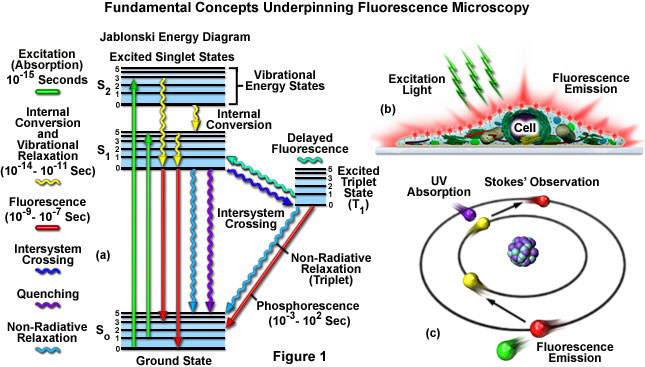
44 fluorescent labels and light microscopy
Fluorescent Nanodiamonds for HeLa Cell Drug Delivery Aug 24, 2022 · The developed fluorescent nanodiamonds were characterized using infrared (IR) spectroscopy, dynamic light scattering (DLS), and secondary electron microscopy. Compared to the free drug with drug release in 12 hours, the fluorescent nanodiamonds functionalized with drug molecules exhibited sustained drug release for over 72 hours. Seeing More with In Silico Labeling of Microscopy Images - Google AI Blog Fluorescence microscopy image of the same cells. The blue fluorescent label localizes to DNA, highlighting cell nuclei. The green fluorescent label localizes to a protein found only in dendrites, a neural substructure.The red fluorescent label localizes to a protein found only in axons, another neural substructure.With these labels it is much easier to understand what's happening in the sample.
Light Microscope- Definition, Principle, Types, Parts, Labeled Diagram ... A light microscope is a biology laboratory instrument or tool, that uses visible light to detect and magnify very small objects and enlarge them. They use lenses to focus light on the specimen, magnifying it thus producing an image. The specimen is normally placed close to the microscopic lens.
Fluorescent labels and light microscopy
Mica | Products | Leica Microsystems No constraints - Select the right modality in real time. Mica unifies transmitted and fluorescence light imaging modalities. You can select from multiple imaging modalities all within one Microhub, including widefield, confocal, THUNDER imaging, LIGHTNING, Z-stacks, time-lapse and more. | ECFP fluorescence is maintained by Glycerol-RI matching in a ... Here, we propose a guideline for 3D light microscopy imaging to achieve single-cell resolution. The guideline includes a validation experiment focusing on five optical clearing protocols. Fluorescence microscopy: established and emerging methods ... - PubMed Numerous practical strategies to enhance fluorescence microscopy experiments are reviewed. The use of instrumentation such as light traps, cameras, objectives, improved fluorescent labels, and image filtration routines applicable to low light level experiments are discussed.
Fluorescent labels and light microscopy. Fluorescent tag - Wikipedia S. cerevisiae septins revealed with fluorescent microscopy utilizing fluorescent labeling In molecular biology and biotechnology, a fluorescent tag, also known as a fluorescent label or fluorescent probe, is a molecule that is attached chemically to aid in the detection of a biomolecule such as a protein, antibody, or amino acid. Novel Fluorescent Label Shines a Light on DNA Structure in Cancer Cells Novel Fluorescent Label Shines a Light on DNA Structure in Cancer Cells March 7, 2022 Researchers have developed a new fluorescent label that gives a clearer picture of how DNA architecture is... Label-free prediction of three-dimensional fluorescence images from ... Although fluorescence microscopy can resolve subcellular structure in living cells, it is expensive, is slow, and can damage cells. We present a label-free method for predicting three-dimensional fluorescence directly from transmitted-light images and demonstrate that it can be used to generate multi-structure, integrated images. Fluorescent Nanoparticles for Super-Resolution Imaging Super-resolution imaging techniques that overcome the diffraction limit of light have gained wide popularity for visualizing cellular structures with nanometric resolution. Following the pace of hardware developments, the availability of new fluorescent probes with superior properties is becoming ever more important. In this context, fluorescent nanoparticles (NPs) have attracted increasing ...
Fluorescence Microscopy vs. Light Microscopy - News-Medical.net This means that fluorescent microscopy uses reflected rather than transmitted light. For example, a commonly used label is green fluorescent protein (GFP), which is excited with blue light and... Fluorescence Imaging - Teledyne Photometrics Fluorescent molecules (known as fluorophores) are used to label samples, and fluorophores are available that emit light in virtually any color. In a fluorescent microscope, a sample is labeled with a fluorophore, and then a bright light ( excitation light) is used to illuminate the sample, which gives off fluorescence ( emission light ). Fluorescence Microscope: Principle, Types, Applications Fluorescence microscopy is a light microscope that works on the principle of fluorescence. A substance is said to be fluorescent when it absorbs the energy of invisible shorter wavelength radiation (such as UV light) and emits longer wavelength radiation of visible light (such as green or red light). Label-free prediction of three-dimensional fluorescence images from ... We present a label-free method for predicting three-dimensional fluorescence directly from transmitted-light images and demonstrate that it can be used to generate multi-structure, integrated...
Saturated excitation microscopy for sub-diffraction-limited imaging of ... The SAX microscopy exploits the saturation fluorescent molecules' excited state population to improve the spatial resolution. 39 - 42 As the SAX is induced by light illumination at a high-excitation intensity, the nonlinear fluorescence signals are localized in the excitation focus spot, resulting in the improvement of spatial in 3-D. Since ... Semiconductor Nanocrystals as Fluorescent Biological Labels Sep 25, 1998 · In addition, nanocrystal probes may prove useful for other contrast mechanisms such as x-ray fluorescence, x-ray absorption, electron microscopy, and scintillation proximity imaging, and the use of far red– or infrared-emitting nanocrystals (InP and InAs) as tunable, robust infrared dyes is another possibility. Site of fluorescent label modifies interaction of melittin with live ... Fluorescence and light microscopy revealed changes in cell morphology after exposure to MLT peptides and showed bleb formation in the plasma membrane of HeLa cells. However, the membrane disruptive effect was dependent upon the location of the fluorescent label on the peptide and was greater when MLT was labeled at the N-terminus. Proline at ... PDF Saturated excitation microscopy for sub-diffraction-limited imaging of ... Keywords: confocal microscopy; fluorescence microscopy; saturated excitation; depth discrimination property; high resolution. Paper 130420RR received Jun. 18, 2013; revised manuscript received Oct. 28, 2013; accepted for publication Nov. 1, 2013; published ... high-image contrast. 1 4 A pinhole placed in front of a light detector is a key ...
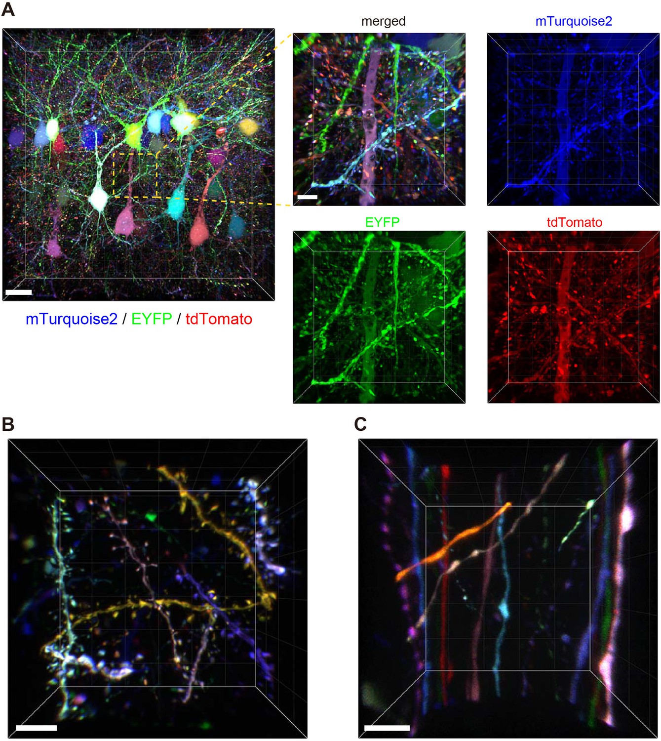
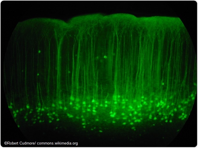
Bright multicolor labeling of neuronal circuits with ...
Fluorescence Microscopy - Explanation and Labelled Images Fluorescence microscopy uses a high-intensity light source that excites a fluorescent molecule called a fluorophore in the sample observed. The samples are labeled with fluorophore where they absorb the high-intensity light from the source and emit a lower energy light of longer wavelength.
ZEISS Microscopy Online Campus | Practical Consideration in ...
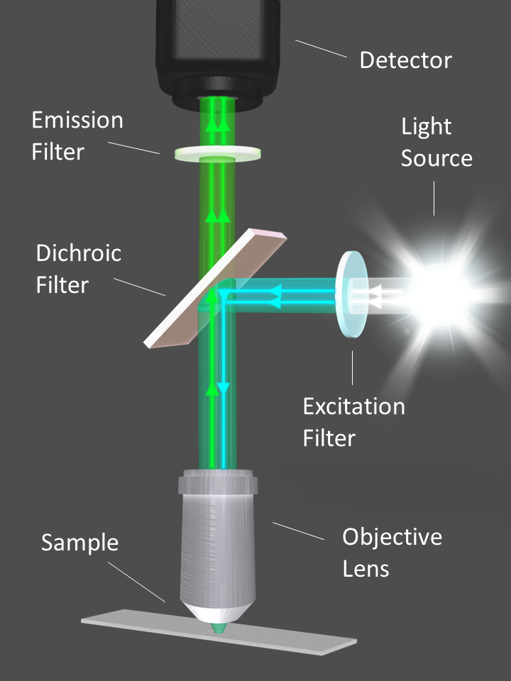
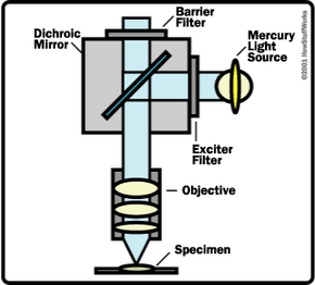
Fluorescence microscope - Wikipedia Typical components of a fluorescence microscope are a light source ( xenon arc lamp or mercury-vapor lamp are common; more advanced forms are high-power LEDs and lasers ), the excitation filter, the dichroic mirror (or dichroic beamsplitter ), and the emission filter (see figure below).

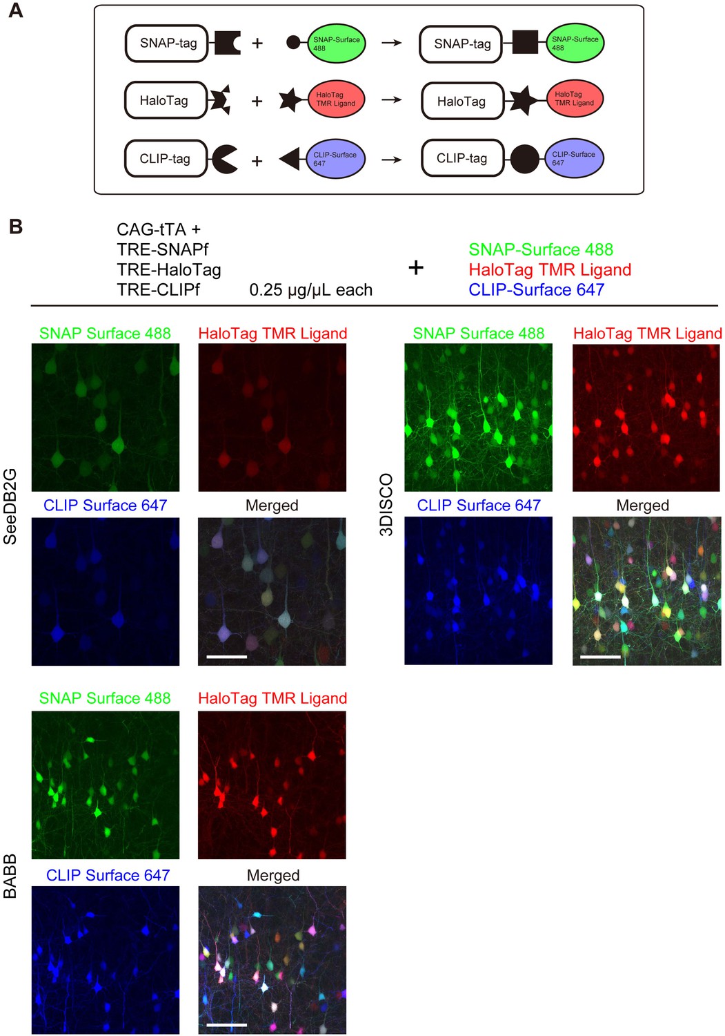
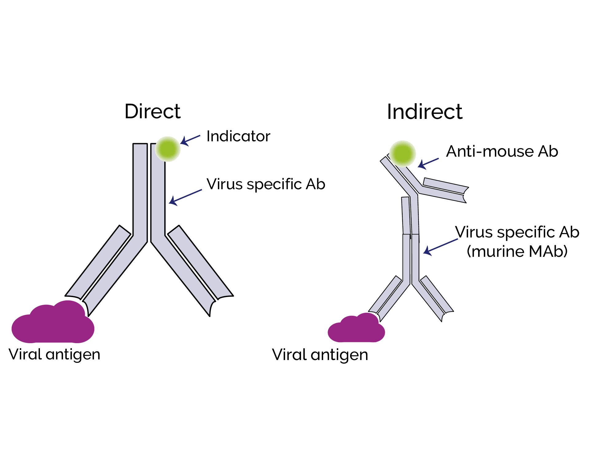
Experimental layout of the multi label fluorescence ...
Fluorescence - Wikipedia A perceptible example of fluorescence occurs when the absorbed radiation is in the ultraviolet region of the electromagnetic spectrum (invisible to the human eye), while the emitted light is in the visible region; this gives the fluorescent substance a distinct color that can only be seen when exposed to UV light. Fluorescent materials cease to ...
Fluorescence Microscopy Virtual Lab
Label-Free Fluorescence Microscopy Through Channels Now, researchers in Sweden have developed a new technique called nanofluidic scattering microscopy, which images the target particles while they are flowing through an optically transparent matrix of nanoscale channels (Nat. Methods, doi: 10.1038/s41592-022-01491-6 ). The technique, which incorporates dark-field light-scattering microscopy ...

Fluorescent microscopy - LNF Wiki
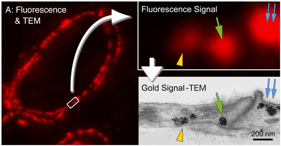
PDF Direct Evidence of Lack of Colocalisation of Fluorescently Labelled ... Fluorescently labelled nanoparticles are routinely used in Correlative Light Electron Microscopy (CLEM) to combine the capabilities of two separate microscope platforms: fluorescent light microscopy (LM) and electron microscopy (EM). The inherent assumption is that the fluorescent label observed under LM colocalises well with the electron dense ...
Bright multicolor labeling of neuronal circuits with ...
Introduction to Fluorescent Proteins | Nikon’s MicroscopyU The brightness and fluorescence emission spectrum of enhanced yellow fluorescent protein combine to make this probe an excellent candidate for multicolor imaging experiments in fluorescence microscopy. Enhanced yellow fluorescent protein is also useful for energy transfer experiments when paired with enhanced cyan fluorescent protein (ECFP) or ...
ZEISS Microscopy Online Campus | Microscopy Basics ...
Introduction to Fluorescence Microscopy • iBiology In this introductory lecture on light microscopy, Dr. Nico Stuurman describes the principles and properties of fluorescence microscopy. Skip to primary navigation; ... 00:01:04;23 so we label microtubules with the fluorescent dye 00:01:08;13 and we have part of the cell labeled in red, 00:01:11;25 and that part is actually the uh, nucleus
Imaging and Microscopy Fluorescence Filters
Caveat fluorophore: an insiders’ guide to small-molecule ... Dec 23, 2021 · From its inception, fluorescence microscopy has been driven by advances in dyes. Early protein labels excited by ultraviolet (UV) light proved difficult to use for cellular imaging, and so Coons ...

Chemosensors | Free Full-Text | “Probe, Sample, and ...
Fluorescence microscopy: established and emerging methods ... - PubMed Numerous practical strategies to enhance fluorescence microscopy experiments are reviewed. The use of instrumentation such as light traps, cameras, objectives, improved fluorescent labels, and image filtration routines applicable to low light level experiments are discussed.
An Overview of Fluorescence Microscopy - Biotium
| ECFP fluorescence is maintained by Glycerol-RI matching in a ... Here, we propose a guideline for 3D light microscopy imaging to achieve single-cell resolution. The guideline includes a validation experiment focusing on five optical clearing protocols.
Hyperspectral multiphoton microscopy for in vivo ...
Mica | Products | Leica Microsystems No constraints - Select the right modality in real time. Mica unifies transmitted and fluorescence light imaging modalities. You can select from multiple imaging modalities all within one Microhub, including widefield, confocal, THUNDER imaging, LIGHTNING, Z-stacks, time-lapse and more.

Label-free prediction of three-dimensional fluorescence ...
ZEISS Microscopy Online Campus | Microscopy Basics ...
Differences between Light Microscope and Electron Microscope
Advanced Light Microscopy Methods - 2008 - Wiley Analytical ...
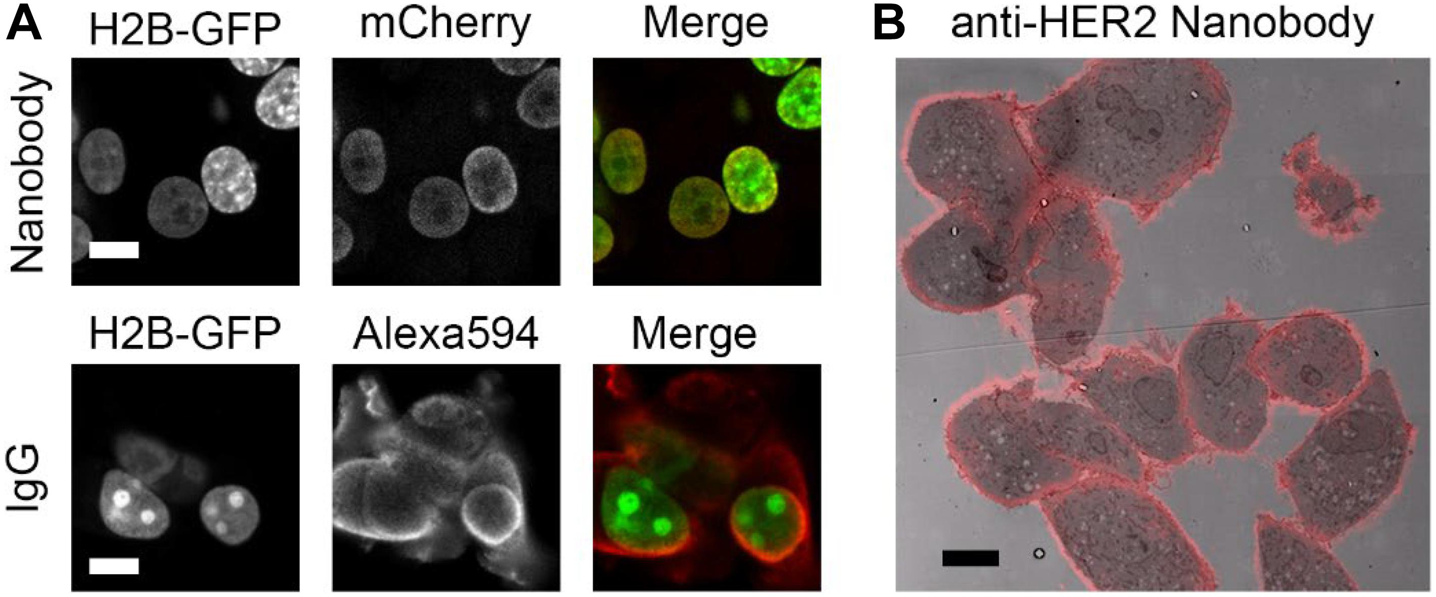
Frontiers | Nanobody-Based Probes for Subcellular Protein ...

Sample Preparation for Fluorescence Microscopy
Fluorescence Microscopy vs. Light Microscopy

Fluorescent Label - an overview | ScienceDirect Topics

Single-cell cytometry via multiplexed fluorescence prediction ...

Fluorescent Microscopy
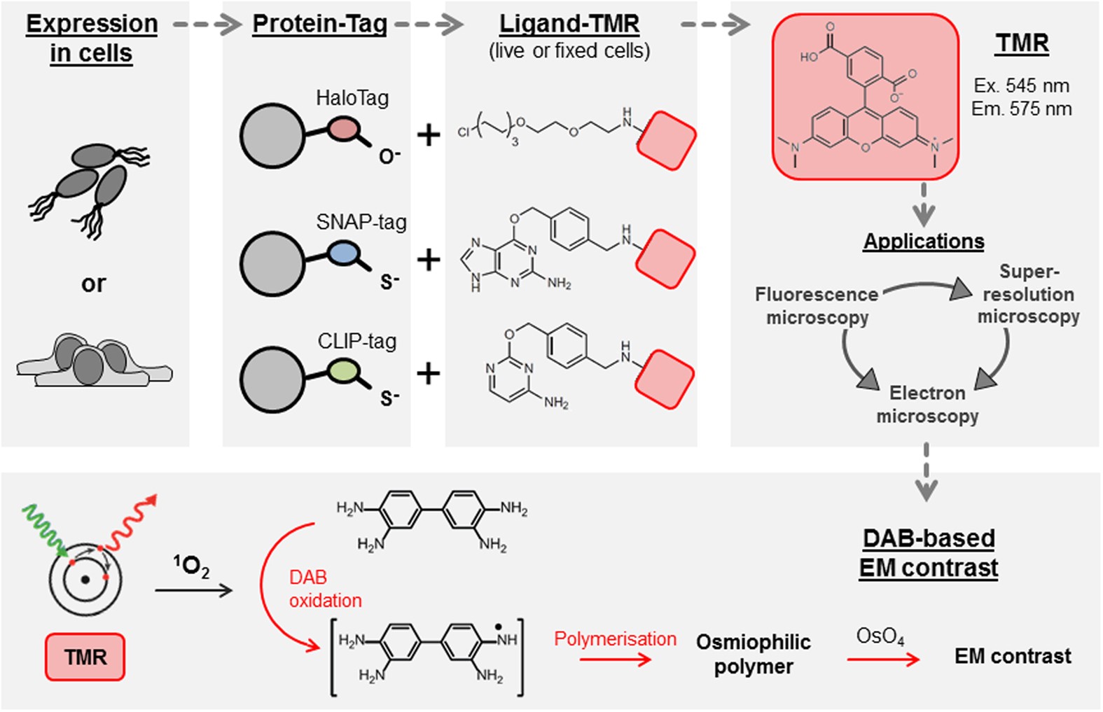
Self-labelling enzymes as universal tags for fluorescence ...
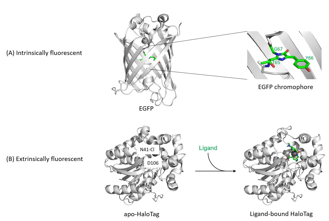
Fluorescent protein tags | Proteintech Group

Fluorescent tag - Wikipedia
Definition > Fluorescence microscope
FluoroNanogold™ combined fluorescent and gold nanoparticle ...
Light Microscopy Techniques in Virology: An Overview

Light-microscopy methods in C. elegans research - ScienceDirect
ZEISS Microscopy Online Campus | Introduction to Spectral Imaging
Fluorescence Microscopy - How Light Microscopes Work ...

Light microscopes — Science Learning Hub

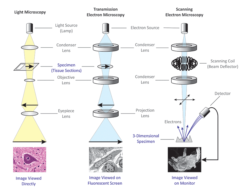
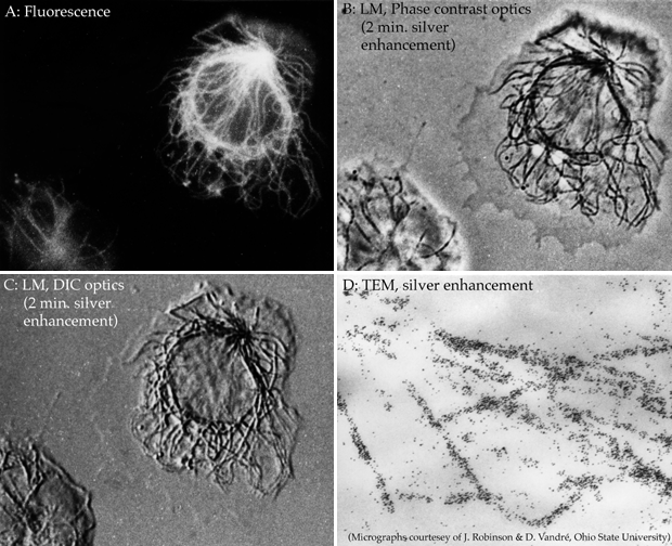
Diagram of labeling used for light and electron microscopy. A ...
FluoroNanogold™ combined fluorescent and gold nanoparticle ...
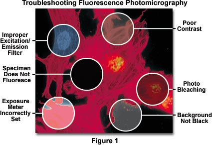
Fluorescence Microscopy Errors | Olympus LS
Dots, Probes and Proteins: Fluorescent Labels for Microscopy ...
The Biological bulletin. Biology; Zoology; Biology; Marine ...
Super-resolution fluorescence microscopy studies of human ...
Correlative fluorescence microscopy, transmission electron ...
A Genetically Encoded Tag for Correlated Light and Electron ...
LABORATORIES 1-6

VIPER is a genetically encoded peptide tag for fluorescence ...

Label-free imaging tool pipeline and application using 3D ...
GFP-tagging in Fluorescence Microscopy
Light Microscopy Techniques Used for Virology- Oxford Instruments
Post a Comment for "44 fluorescent labels and light microscopy"