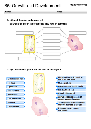
42 onion cells under microscope with labels
Onion Epidermis - kuensting.org Onion epidermal cells, iodine stain, 400X. The nucleus of an onion epidermal cell, 1000X magnification. ... Required practical - using a light microscope - Cells in ... - BBC Bitesize Record the microscope images using labelled diagrams or produce digital images. When first examining cells or tissues with low power, draw an image at this stage, even if going on to examine the ...
Microscope Cell Lab: Cheek, Onion, Zebrina - SchoolWorkHelper The first lab exercise was observing animal cells, in this case, my cheek cells. The second lab exercise was observing plant cells, in this case, onion epidermis. The third lab exercise was observing chloroplasts and biological crystals, in this case, a thin section from the Zebrina plant. The first thing that was done in this lab exercise was ...
Onion cells under microscope with labels
Observing Onion Cells under a Microscope - Blog, She Wrote you'll need to stain the onion cells before you observe them under the microscope. There are different types of stains depending on what type of cell you are going to look at. Iodine - dark stain that colors starches in cells. In an onion cell, it will make the cell wall more visible. It provides some contrast for viewing under a microscope. Looking at the Structure of Cells in the Microscope Both types of light microscopy are widely used to visualize living cells. Figure 9-7 Two ways to obtain contrast in light microscopy. (A) The stained portions of the cell reduce the amplitude of light waves of particular wavelengths passing through them. A colored image of the cell is thereby obtained that is visible in the ordinary way. (more...) Onion Root Tip Mitosis - Stages, Experiment and Results · Cover the sample (root tip) with a coverslip and gently press the coverslip down, then examine the slide under the microscope starting with low magnification * For this experiment, a properly prepared slide should appear light pink due to the stain to almost colorless. * Unused roots can be stored in 70 percent alcohol. Results
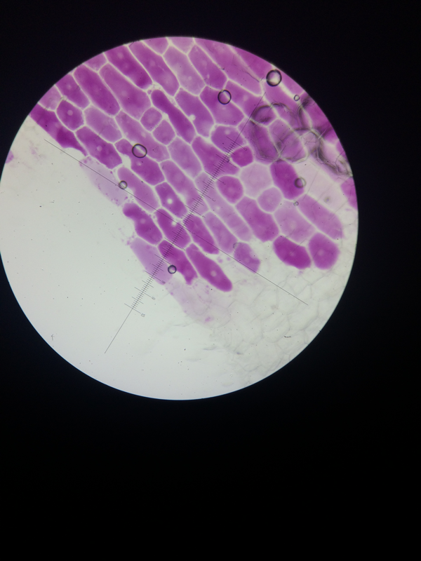
Onion cells under microscope with labels. animal cell under microscope 100x - Lanora Vanmeter The animal cell structure is the most prominent in human cheek cells. The granulated area is the cell Cytoplasm while the huge round part is the Nucleus. Firstly when a slide of onion root cell plant cell and whitefish cell animal cell were observed under a microscope at 40X magnification nucleus were able to be identified. Animal Cell Diagram Under Microscope Labeled Animal Cell Diagram Under Microscope Labeled Sunday, April 18th 2021. | Diagram Animal Cell Diagram Under Microscope. Function cell does in the body dictate the change and adaptation done by cell. When observing onion cells, there is the Cell Surface Membrane which is present in all living cells. ONION CELLS VIDEO - YouTube Video shows how to make a wet mount slide to view onion cells under the microscope. Biology Experiment Examination of Onion Cell in Light Microscope Place the single layer of onion cell epithelium on a glass slide. Make sure that you do not fold it over or wrinkle it. Place a drop of iodine stain on your onion tissue. Put the cover slip on the stained tissue and gently tap out any air bubbles. Observe the cells under 4x, 10x, and 40x with the diaphragm wide open.
Labeled Onion Cell Under Microscope 40x - Micropedia For the experiment you will only need onion dropper and the microscope container and tools are optional. Labeled onion cell under microscope 40x. While photosynthesis takes place in the leaves of an onion containing chloroplast the little glucose that is produced from this process is converted in to starch starch granules and stored in the bulb. Onion Peels Observed Under the Microscope - First-Learn.com But if it is observed under microscope in high resolution then presence of cell vacuoles can be observed properly. Characteristics features of the onion peels are -. 1. Cells are firmly bound to each other. 2. Nucleus present in the cells are slightly towards the periphery of the cells. Which is the one of the confirmation point of onion peel. PDF Onion Cells - Investigation - Exploring Nature 5. Observe the onion tissue under the microscope at 4x, 10x and 40x with lots of light (open diaphragm). Then slowly close the diaphragm while observing the image to find the best light for seeing cellular details. 6. Draw a section of onion skin cells at 10x magnification. Then switch to 40x and draw one cell and label it. Questions: 1. Onion Cell Lab Report.docx - Onion Cell Lab Report By station, remove the single layer of epidermal cells from inner side of the scale leaf. 3(Place the single layer of onion on a glass slide. 4(Place a drop of iodine stain on your onion tissue. 5(Put the cover slip on the stained tissue and gently tap out any air bubbles. 6(Observe the cells under the microscope and see you results.
Onion Epidermal Cell Labeled Diagram - schematron.org Draw a labelled diagram of an onion epidermal cell seen under the microscope. ( 4 marks) e The onion epidermal cells are not green in colour because they lack. The epidermal cells of onions provide a protective layer against viruses and fungi that may harm the sensitive tissues. PDF Onion Cell Lab - SomeWaresInMaine 1. Onion layer (tissue) 2. iodine stain 3. slide & cover slip Procedure: 1. Carefully separate the thin film tissue from between two layers of an onion 2. Carefully place a small sample of this tissue onto a slide - avoid folds & creases 3. Put a drop of iodine stain on the tissue 4. Carefully place a coverslip to avoid air bubbles 5. Onion Cell Under Microscope 400x Labeled - Micropedia Onion Epidermis. Px12 016a Plant Stomata Guard Cells Opened Zebrina Spp 400x. Onion Cells Under The Microscope Requirements Preparation And. Microscopy Biology Libretexts. Cells Under A Microscope By Jaimarie Nelson. Cell Lab. Microscope Lc Lessons Tes Teach. Human Cheek Cell Hamle Rsd7 Org. Animal Cell Under Light Microscope Labelled : Draw and label the ... Onion cell diagram labeled structure of animal cell and plant cell under microscope. An organelle found in large numbers in most cells, in which the biochemical processes of respiration and energy production occur. Under a light microscope, the cell membrane, nucleus and cytoplasm of a cheek cell (animal cell) can be observed.
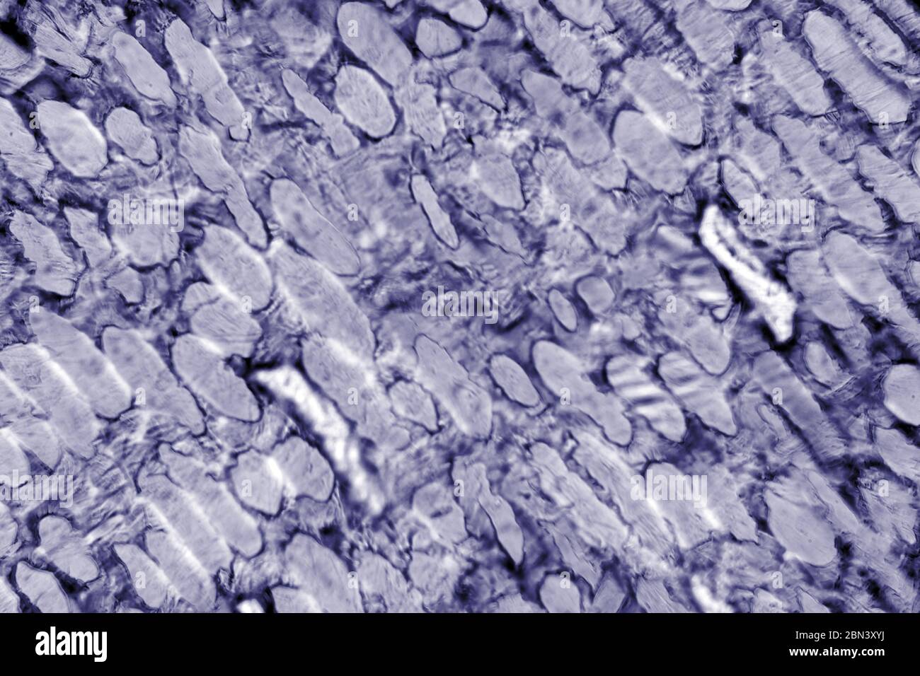
Onion Cells High Resolution Stock Photography and Images - Alamy
What organelles are in an onion cell? - Biology Stack Exchange You cannot see most of these as they appear translucent as well as being too small to see under the light microscope. You need an electron microscope to view these. Note: chloroplasts are not present in an onion cell as it is not a photosynthesising cell. This is a typical onion cell slide with labels:
Biology Pictures: Onion Cells under Microscope
DOC The Onion Cell Lab - chsd.us Place the single layer of onion cell epithelium on a glass slide. Make sure that you do not fold it over or wrinkle it. Place a drop of iodine stain on your onion tissue. Put the cover slip on the stained tissue and gently tap out any air bubbles. Observe the cells under 4x, 10x, and 40x with the diaphragm wide open.

Cell structure | Cells as the basic units of life | Siyavula
animal cell under microscope labeled - Mad Thing Blogging Galleria Di ... Onion Epidermis Under Light Microscope Purple Colored Large Cells Project Microscopic Photography Epidermis. Within the cell there is a shape of round with a circular structure of granulated part on the. ... Animal Cell Diagram Under Microscope Labeled. Add a drop of purple stain specific for animals and cover with a cover slip. Draw a diagram ...

swifty science: onion cell lab
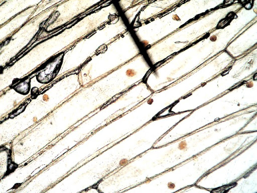
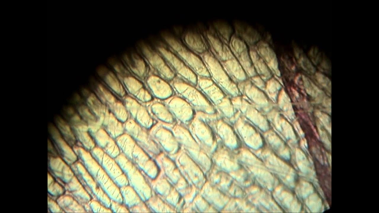
Onion Skin Epidermis Sample under microscope 4x,10x Magnification A sample on an onion skin epidermis diyed in blue for visibility, viewd under the microscope at 4x and 10x magnification.microscope:Biolux model :AL
Red Onion Cell Under Microscope Labeled - Micropedia
Onion Cells Under a Microscope - Requirements/Preparation/Observation Add a drop of iodine solution on the onion membrane (or methylene blue) Gently lay a microscopic cover slip on the membrane and press it down gently using a needle to remove air bubbles. Touch a blotting paper on one side of the slide to drain excess iodine/water solution, Place the slide on the microscope stage under low power to observe.

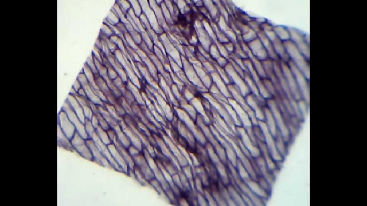
Biology 1 - Onion cell microscopy - YouTube
animal cell under microscope labeled - Rayford Runyon When observing onion cells there is the Cell Surface Membrane which is present in all living cells. Most cells both animal and plant range in size between 1 and 100 micrometers and are thus visible only with the aid of a microscope. While observing with tissues or on tissue.
Onion cells under the microscope: 40X - 100X - 400X - YouTube
DOC Plant and Animal Cells Microscope Lab - hillsboro.k12.oh.us Make a drawing of one onion cell, labeling all of its parts as you observe them. (At minimum you should observe the nucleus, cell wall, and cytoplasm.) Cheek cells 1. To view cheek cells, gently scrape the inside lining of your cheek with a toothpick. DO NOT GOUGE THE INSIDE OF YOUR CHEEK! (We will observe blood cells in a future lab!!) 2.
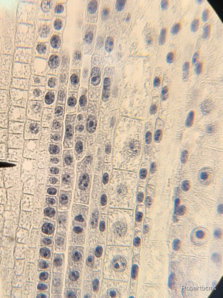
"Biology- onion root tip cells under microscope" iPhone Case & Cover by Robertbcms | Redbubble
Onion Skin Cells - Investigation - Exploring Nature 5. Observe the onion tissue under the microscope at 4x, 10x and 40x with lots of light (open diaphragm). Then slowly close the diaphragm while observing the image to find the best light for seeing cellular details. 6. Draw a section of onion skin cells at 10x magnification. Then switch to 40x and draw one cell and label it.
Cells: Microscope cheek and onion cell | Teaching Resources
Observing Onion Cells Under The Microscope Afterwards, carefully mount the prepared and stained onion cell slide onto the microscope stage. Make sure that the cover slip is perfectly aligned with the microscope slide, and that any excess stain has been wiped off. Secure the slide on the stage using the stage clips.
Light Microscope Onion Cell Labeled - Micropedia
vii Sketch the onion peel cell as seen under the microscope Label the ... Put a drop of water in its centre and transfer the peel from the petridish to the slide with the help of a brush. Place the coverslip. (viii) Remove the extra water by placing the slide within a folded filter paper. (ix) Examine the slide first under low power and then under high power. (x) Record your observations.
Onion Cells Under Microscope! REALLY COOL!!! - YouTube
Microscopy, size and magnification - Microscopy, size and ... - BBC Place cells on a microscope slide. Add a drop of water or iodine (a chemical stain). Lower a coverslip onto the onion cells using forceps or a mounted needle. This needs to be done gently to...

Cells Zone – I'm a Scientist, Get me out of Here!
Onion Root Tip Mitosis - Stages, Experiment and Results · Cover the sample (root tip) with a coverslip and gently press the coverslip down, then examine the slide under the microscope starting with low magnification * For this experiment, a properly prepared slide should appear light pink due to the stain to almost colorless. * Unused roots can be stored in 70 percent alcohol. Results

How to Observe Onion Cells under a Microscope | Things under a microscope, Science cells, High ...
Looking at the Structure of Cells in the Microscope Both types of light microscopy are widely used to visualize living cells. Figure 9-7 Two ways to obtain contrast in light microscopy. (A) The stained portions of the cell reduce the amplitude of light waves of particular wavelengths passing through them. A colored image of the cell is thereby obtained that is visible in the ordinary way. (more...)

Fanos' MCB Blog: Onion Skin
Observing Onion Cells under a Microscope - Blog, She Wrote you'll need to stain the onion cells before you observe them under the microscope. There are different types of stains depending on what type of cell you are going to look at. Iodine - dark stain that colors starches in cells. In an onion cell, it will make the cell wall more visible. It provides some contrast for viewing under a microscope.

Rens blog : Science, cells
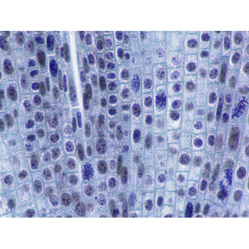
Slide, Microscope, Onion Root Tip Mitosis
Post a Comment for "42 onion cells under microscope with labels"